|
HALE
VETERINARY CLINIC |
|
|
| Oral Exams
| Periodontics | Endodontics | Restorations | Prosthodontics | Orthodontics Tumor Removal | Facial Fracture Repair| Radiography | Anesthesia & Radiography | Juvenile Dentistry | Cats |
|
HALE
VETERINARY CLINIC |
|
|
| Oral Exams
| Periodontics | Endodontics | Restorations | Prosthodontics | Orthodontics Tumor Removal | Facial Fracture Repair| Radiography | Anesthesia & Radiography | Juvenile Dentistry | Cats |
| For Veterinarians | We offer an instrument sharpening service to veterinary
facilities as outlined in this brochure - instrument sharpening
services. Please call to schedule your instruments arrival. Turn
around time (from when the instruments leave your office until you have
them back) will be at least one week. Dr Hamilton is happy to review case histories, photos and radiographs from referring veterinarians as a professional courtesy. To access this service, complete Step One and Step Two on our landing page and send the necessary information to info@toothvet.ca He would also encourage general practitioners to post their questions on the Dental Discussion Folder at www.vin.com to get a range of opinions and options and to help others learn from the discussion.
|
| For Owners | We offer a wide range of dental and oral services as outlined below. |
|
|
Introduction Dr. Hamilton provides a wide range of dental and oral services for dogs and cats. In the bad old days, little attention was paid to pet’s teeth. They were largely ignored until they were seriously loose and then they were extracted. Over the past few decades, pet animals have taken on an increasingly important role in people’s lives. As a result, owners and veterinarians came to realize that quality dental care could provide the same benefits for pets as it does for humans. Early detection and treatment of dental and oral problems can not only save teeth, it can also dramatically improve the quality of life. Think about it from the pet’s point of view. They use their mouths to eat, as we do. But they also use their mouths in much the same way we use our hands – for work and play and to interact with the world around them. When their mouths are diseased or injured, it is as painful for them as it would be for us. Competition and natural selection have ‘taught’ them to carry on bravely and keep eating, despite the pain, but you can rest assured that dental pain in pets is very real. Regardless of what problems you pet may be having, Dr. Hamilton's #1 priority is always to provide and maintain a mouth free of pain and infection/inflammation. Nothing is more important than this. Here are some of the dental services provided by Dr. Hamilton. |
 |
Oral Examinations All work done by Dr. Hamilton starts the same way – with a referral from a general veterinary practitioner (either at their suggestion or at the owner’s request). After a review of the pertinent medical history and a telephone consultation, an appointment is made and the pet is examined at our Guelph office. Early on in the visit, Dr. Hamilton will do as thorough an oral examination as the patient will allow. This pre-operative examination allows Dr. Hamilton, in direct consultation with the owners, to develop a tentative treatment plan and an itemized estimate of the cost. If the owners consent to proceed, the animal is anesthetized and a much more detailed oral examination, with dental radiographs (x-rays) is carried out. The treatment plan and estimates then are revised as needed in light of any new findings, again in direct consultation with the owners. Treatment of the problem(s) follows immediately. Therefore, diagnosis, treatment planning and treatment are all provided in one visit. The animal is typically discharged within an hour of the end of the procedure. |
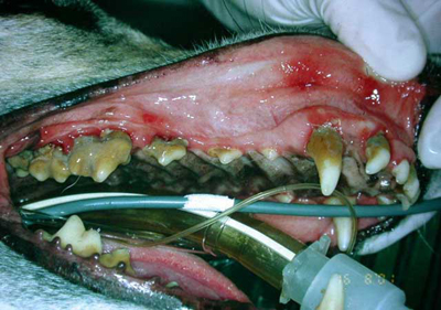 |
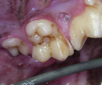
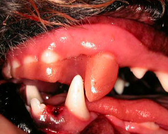
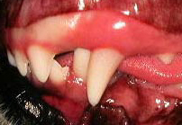
Periodontics Periodontics deals with the tissues that support the teeth – gums, bone and periodontal ligament. The most common disease in pet animals is periodontal disease – it affects about 90% of dogs over five years of age and a similar number of cats. Periodontal disease often starts as simple gingivitis (inflammation of the gums), which is reversible if treated early. If left alone, gingivitis will often progress to involve gum recession, periodontal bone loss and pocket formation. Eventually, the teeth may fall out, but that takes years. In the mean time, aggressive bacteria are allowed to enter into the bloodstream through the diseased periodontal tissues. There is an ever-growing body of research showing that chronic dental infection can have serious negative impact on the patient's health. Prevention is the best approach, but once periodontal disease is established, appropriate treatment can often save teeth and reduce the risk of other serious health problems. For lots more information, see the Periodontal Disease section on the Old CUSP Articles page. |

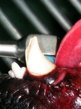
And the final appearance of the tooth. |
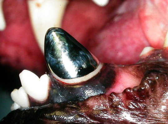
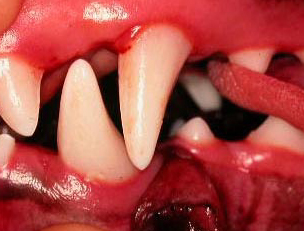
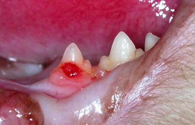
Endodontics Endodontics involves treatment of the pulp of the tooth – the nerves, blood vessels and other soft-tissue inside the tooth. Animal teeth are subject to wear and fracture from playing, chewing and accidental trauma. When the crown (visible part) of the tooth is damaged (fractured or worn down), the pulp often becomes exposed to bacteria. The result is infection and death of the pulp and then infection extends through the root tip(s) into the surrounding bone. For teeth with endodontic disease, there are two options only – either extraction of the tooth or some form of root canal treatment. Leaving an endodontically diseased tooth alone is not an option. For very recent fractures (less than two days) in young animals (less that 18 months), it is often possible and desirable to keep the pulp alive and healthy by removing just a portion of it and then sealing the tooth to prevent bacterial invasion of the remaining pulp. In older injuries and older animals, it is usually wisest to do a full root canal treatment (remove all of the pulp and fill in the entire pulp chamber). After root canal treatment (and often before it as well), the tooth is dead on the inside, but it can still remain in the mouth and be perfectly functional and comfortable for many, many years. Sometimes a tooth will suffer an injury that does not fracture the crown, but that still causes inflammation and death of the pulp. These teeth may be recognizable by the discolouration of the crowns. Teeth that are pink, gray or tan in colour (not just staining on the surface), have dead pulps and also require extraction or root canal treatment. Otherwise, chemicals leaking from the dead pulp cells will ooze out through the root tip and cause inflammation in the periodontal ligament space and surrounding bone. This is not only chronically painful, but also provides a safe haven for bacteria that may be circulating in the blood-stream. For lots more information, see the "Endodontic" section on the Old CUSP Articles page. |

Left
untreated it could have progressed to this! |
Restorations Though cavities are not as common in dogs as they are in humans, they do occur. When they occur, they are treated in exactly the same way. The diseased portion of the tooth is removed and the defect filled with a restorative such as a bonded composite restorations. When a tooth has had root canal work done, there is a hole (or a few holes) in the crown used to gain access to the inside of the tooth. These holes are also filled with bonded composite resins. Dogs may also have congenital defect of the enamel on the crowns of the teeth. Not only unsightly, these defects can expose the underlying dentin, which is sensitive. Bonded restoration make the teeth smoother so they look better and are easier to keep clean, but more importantly, they act as a bandage to protect the sensitive pulp tissues inside the tooth. For more information on caries (cavities) in dogs, see the article in the Other Pathology section of the Old CUSP Articles page. |
|
Prosthodontics
In cases where a dog has fractured/damaged the crown of a tooth due to some long-standing habit or occupation, following root canal treatment, it may be important to protect the tooth from further damage. This is may be achieved by covering the remaining natural crown with a cast metal crown. It is important to keep in mind that a cast metal crown such as this only protects the tooth it is covering. If the patient continues with the behaviour that broke this tooth, he is likely to break more teeth. Therefore, it is even more important to alter behaviour and habits if at all possible to protect all of the teeth. The decision whether or not to place a cast metal crown is based on consideration of many factors, well beyond the scope of this small comment box. To be honest, it is my opinion that there are very few cases in which a cast metal crown is indicated or appropriate. |
|
Orthodontics It has been said that not every animal needs a perfect bite, but all deserve a comfortable bite. Many pets develop perfectly normal and healthy bites (that is, their teeth and jaws all fit together properly). Some animals, however, develop problems as they grow. One jaw may be too long or too short, one or more teeth may be out of place or crowded. In cases where these orthodontic problems are causing abnormal tooth-to-tooth or tooth-to-soft tissue contact something needs to be done. Treatment options may include selective extraction, crown height reduction or use of an orthodontic appliance to move teeth to an acceptable position. Each case must be evaluated very carefully to determine the right course of action, considering the problem, the animal’s personality and the owner’s expectations. Treating orthodontic problems raises some interesting ethical concerns. Many orthodontic problems are genetic and can be passed on to future generations. Therefore, affected animals should be neutered to prevent the passing on of the "bad" genes. Some show-dog owners, looking to maximize their chances in the ring may request orthodontic correction. To make an abnormal dog look normal for the show ring is fraud and Dr. Hamilton will have nothing to do with this. |
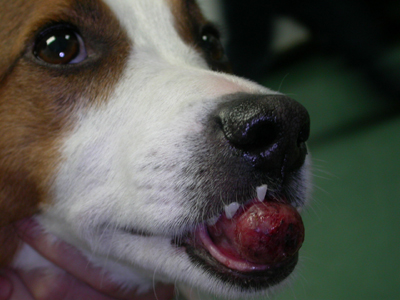 This dog had a large malignant tumor of his lower jaws as seen in the photo above and x-ray below 
|
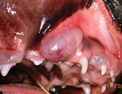
Tumor
Removal Oral growths are a fairly common finding. They may be benign such as gingival hyperplasia or malignant cancer such as squamous cell carcinoma. Even benign growths can cause serious local problems due to the space they occupy in the mouth. For the vast majority of oral masses, treatment is based on early and aggressive surgical removal of the mass with microscopic evaluation of the removed tissue by a veterinary pathologist. While it may be upsetting for owners to learn that their pet requires surgery to remove tumor, teeth and bone, quality of life after surgery is typically very good. Dogs (particularly) and cats often tolerate these surgeries very well and adapt to eating and drinking without trouble. While some tumors can provide insurmountable challenges, many can be managed well. The keys include early detection, accurate assessment/diagnosis and rapid, aggressive action to remove the mass before it grows larger or spreads to other part of the body. |
 This dog had a fracture of his left mandible just in front of the 1st molar.
|
Facial
Fracture Repair Trauma to the head can result in fractures to the upper and/or lower jaws. As animals need to continue to eat and drink, it is important to stabilize these fractures quickly and in a way that allows the patient to continue to use their mouth right away. It is also important to restore proper anatomic and occlusal alignment and to avoid causing damage to the remaining teeth. There are various strategies available and each case needs to be assessed on its own merits to decide what technique(s) to use. For more on fracture repair, see the Oral Trauma Section on the Old CUSP Articles page. |
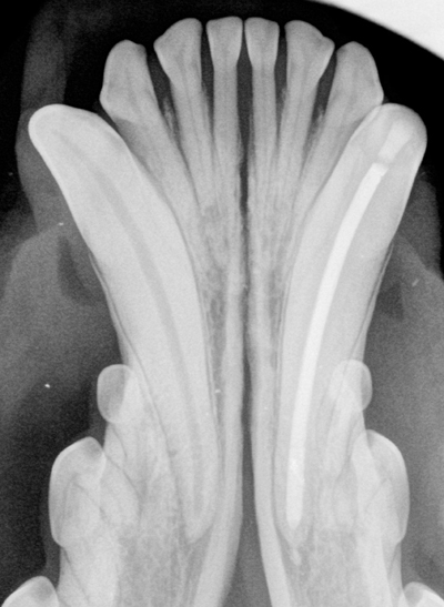
 A normal radiograph of the back of the right lower jaw in a dog. |
Radiography Radiographs (X-rays) are an essential diagnostic tool in dentistry and oral surgery. Much of the tooth lies hidden below the gums and encased in bone. The only way to accurately assess the root structure and surrounding bone is with intra-oral dental radiographs. We use small, flexible sensor plates, placing them right in the mouth to get detailed images of the area in question. Getting diagnostic radiographic images is just step one. The next step is to accurately evaluate that image and know what it means. Dr Hale is often called upon by colleagues to help with interpretation of the images they have obtained. For more on intra-oral dental radiography, see that section on the Old CUSP Articles page. |
|
|
Anesthesia Virtually all dental and oral work requires that the patient be anesthetized. Even the detailed examination and radiographs (X-rays) require anesthesia. However, advances in anesthesia (drugs, monitoring, life-support…) have made general anesthesia a very reasonable risk for the huge majority of pets. Certainly we do not want to give an anesthetic for no reason, but if there is a dental or oral problem that requires attention, anesthesia is an invaluable tool. For more on the subject of the need for general anesthesia for dental patients, see PDFfiles/CVO_position.pdf. As well as general anesthesia, we also employ local anesthetic nerve blocks during surgical procedures as part of the pain management protocol. For more on this, see PDFfiles/LocalAnesthesia.pdf. |
|
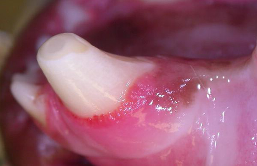
Juvenile
Dentistry The mouths of young animals should be watched closely from a young age by both the owners and the veterinarian. If deciduous (baby) teeth get broken, they should be immediately removed, because leaving them in place will allow the pulp (soft tissue inside the tooth) to become infected and the infection can then spread into the jawbone. As well as being painful, this can also cause damage to the permanent teeth that are forming inside the bone near the root of the deciduous teeth. Orthodontic problems usually develop as the animal is growing. Catching the problem early and taking appropriate steps can often reduce the significance of the problem. Deciduous teeth that do not fall out on time should be removed right away. If the adult tooth comes in and the deciduous tooth is still in place, there are two teeth occupying space meant for one tooth. This causes crowding and can lead to all sorts of problems. For lots more information, see the Developmental Problems section of the Old CUSP Articles page |
|
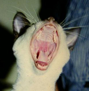
Cats While cats are prone to all the same problems as dogs (malocclusion, tumors, traumatic dental and facial fractures, periodontal disease), they also have a few problems for which they are particularly known. Tooth resorption is a situation in which the animal's own cells eat away at the roots of the teeth. We do not know for sure what causes this condition and we have no recommendations currently to prevent this painful and destructive condition. All we can do is extract affected teeth, monitor for new lesions and deal with them as they arise. By the way, we are starting to find tooth resorption affecting dogs quite commonly as well. Another very frustrating and painful feline condition is one that has a number of names but which I refer to as chronic oral inflammatory disease. There is a great deal we do not yet know about this condition but for the moment, what we do know is that the best (possibly only) hope that these cats have for lasting and meaningful relieve is whole-mouth extraction. The sooner this surgery is done, the better the prognosis. Trying to manage this condition while there are any teeth or roots left in the mouth is doomed to failure and can often cause more problems from the side-effects of the drugs given. For more on these subjects, see the Feline Issues section of the Old CUSP Articles page. |
| Top of this page | |
|
|
519-822-8598 © 2010 Hale Veterinary Prof. Corp. |